However, to ensure such positive outcomes, it that the sponsor or CRO companies should choose the right imaging partner.
FDA & Imaging in Clinical Trials
In the year 1997, the Food and Drug Administration Modernization Act (FDAMA), opened the door for imaging modalities to be used as a product development tool in pharmaceutical or medical device clinical trials. This allowed the inclusion of imaging data (generated through imaging modalities) for regulatory submission. In August 2011, FDA released Clinical Trial Imaging Endpoint Process Standards which described standards that sponsors should use to ensure that clinical trial imaging data is obtained in a manner that:
-
- It complies with trial protocol and standards,
- It maintains imaging data quality within and among clinical sites
- It provides a verifiable record of the imaging process
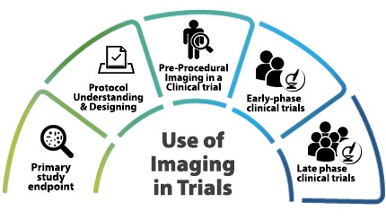
Use of Imaging in Trials
A recent report showed “The global imaging clinical trial market size was $773.4 million in 2016 and is poised to grow at a CAGR of 6 to 8 percent between 2016 and 2020”.

1. Primary study endpoint
Using imaging as a primary, surrogate, and quantitative biomarker has been proven to accelerate clinical trials that have helped bring new drugs to market sooner for various unmet needs (Orphan drugs etc.) However, forethought should be exercised during clinical trial study design and when using imaging as an endpoint.
2. Protocol Understanding & Designing
Every clinical trial begins with the development of a clinical protocol that is compliant with regulatory/GCP requirements. When imaging is added as an endpoint in clinical trials, the right guidance for the right protocol design plays an important role in assisting the sponsor for faster drug approval. Such guidance requires vast knowledge and experience and focuses on on-site training, image acquisition, interpretation, archival, quality assurance, adjudication, EDC submission etc. It is wise to involve an imaging partner in protocol design since it helps include investigational site feasibility assessment and availability of specific imaging hardware and software.
3. Pre-Procedural Imaging in a Clinical trial
Clinical trials perform various screening tests to assess whether prospective subjects are appropriate candidates and if they fulfill the criteria for inclusion in the study. Non-invasive cardiac imaging is one of the extensively used imaging procedures as a pre-procedural patient selection technique.
4. Early-phase clinical trials
Early-phase trials typically present less logistical challenges, and hence imaging is a natural fit for early phase trials.
5. Late phase clinical trials
In the later phase of a Clinical trial, imaging focus shifts toward efficient trial management by streamlined operational workflow for easy regulatory approval.
While on the one hand there are numerous benefits of incorporating imaging into clinical trials, on the other hand there exist unique challenges.
- Need for Subspecialists: A given trial can have a variety of modalities that require review by clinical expert readers. For examples in oncology clinical trials, therapy is assessed using imaging response criteria involving imaging biomarkers. Various Imaging response criteria are used to determine the endpoints. Imaging biomarkers are characteristics extracted from images under study and may vary with a tumour or cancer types and with imaging modalities. Response criteria are created using specific imaging biomarkers with metrics and sets of rules, to provide accurate and reproducible measurements of tumours during therapy
- In various multicentric clinical trials, due to a shortage of trained staff and the lack of a cloud base platform, establishing the right imaging technique and seamless image acquisition become a challenge. While the lack of right imaging technique leads to suboptimal image capture; delay in image acquisition leads to delayed interpretation etc.
- Lack of high-speed network connections at trial sites can hinder the quick transfer of images and lead to image latency.
- Lack of software systems on the market that can effectively document the rationale for changes or modifications in clinical trials (audit trail) to meet 21 CFR Part 11 compliance.
- Lack of peer review and lack of stringent quality assurance process leading to a difference in evaluation and assessment.
- Lack of centralized data collection: A lack of centralized data collection and assessment results in bias in the reading of images
Considering all of these it becomes a mandate to choose the right core partner for successful outcomes. The imaging partner should be able to support the clinical trial in any geographic location, and should have
- A large pool of expertise,
- Be capable of providing strategic advice on biomarkers,
- Assist in protocol development and
- Demonstrate how medical imaging endpoints can provide objective evidence of a drug’s safety and efficacy.
Top reasons Why Image Core Lab is your Right Imaging partner

Our Therapeutic expertise
A cohesive pool of globally accredited radiologists (ABR& FRCR) provides an innovative environment for a broad range of Image Interpretation Scenarios. We ensure that the right radiologist provides the right report. Whether it is a breast or ovarian cancer trial; Cardiac or Neurological trial; Lung nodule assessment or Airway assessment, we have the optimal expertise for your trial imaging.
1. Oncology
Oncology trial is currently ICL’s fastest growing therapeutic area with over 50% of engagements in oncology trials. Experienced in large cancer trials with irRC and RECIST 1.1 protocols. Recently, RECIST and RECIST 1.1 were done together in the same trial.
2. Neurological
Strategic and practical support from experts that will boost your trial process and simplify your product development process. Our fellowship-trained neuroradiologists bring in a subspecialty level experience on neurodegenerative diseases such as Alzheimer’s Disease and Parkinson’s Disease, demyelinating disorders, stroke, epilepsy, neuro-oncology and post-traumatic disorders.
3. Cardiac and vascular
Our strength in Cardiac MR (CMR) image analysis, includes Anatomic Perfusion, Functional Imaging, Graft Patency and Stent Studies. Our expertise in Nuclear Cardiology further complements these services. A global team of in-house cardiovascular experts, who are highly experienced in image analysis of clinical research and drug development provide high-quality interpretation services.
4. Thoracic and pulmonary imaging
We provide the highest level of expertise in the evaluation of chest radiographs and high-resolution CT of pulmonary disorders, including interstitial lung disease, chronic obstructive pulmonary disease, occupational lung diseases, pulmonary thromboembolism, bronchopulmonary infections and tumours.
CLINSpa– Technology Platform
21 CFR Part 11 compliant Technology enabled radiology practice
Encrypted and redundant network compliant with HIPAA standards.
CLINSpa’s tried and tested end to end imaging trial workflow advances the client’s imaging trial process and helps align the imaging trial timelines with overall developmental goals. A platform that is convenient and exclusively designed to cater to all your imaging trial workflow needs.
To find out more about our Services, or how we can help you manage imaging requirements of your trial talk to us today.

