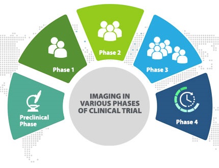
Imaging techniques are increasingly used in oncological clinical trials to provide evidence for decision making. The conventional morphological imaging techniques and standardized response criteria based on tumour size measurements are used increasingly for defining key study endpoints. Non-invasive imaging using computed tomography (CT), magnetic resonance imaging (MRI) and fluorodeoxyglucose (FDG) positron emission tomography (PET)/CT plays a significant role in generating primary, secondary and exploratory study endpoints. In later stages of oncological drug development, imaging forms the basis of robust response and progression criteria to interrogate the drug in a large number of clinical trial subjects.

Preclinical Phase
The primary objective of the preclinical phase of drug development is to establish the safety profile of the drug molecule before clinical testing. This phase evaluates of mutagenicity, carcinogenicity, teratogenicity, pharmacokinetic and pharmacodynamic properties of the molecule that provides insight into the in vivo properties before initiating clinical investigations.
Imaging techniques such as CT, MRI (including functional imaging techniques such as magnetic resonance spectroscopy (MRS), dynamic contrast-enhanced MRI (DCE-MRI), diffusion-weighted MRI (DW-MRI), PET imaging and ultrasound may be used for this. In the preclinical trial stage these may need to be performed in the laboratory setting.
Phase I Clinical Trial
This phase of clinical trial aims at evaluating drug pharmacokinetics and pharmacology across a range of drug doses. In oncology Phase I trials, subjects typically have refractory advanced solid tumours.
Imaging such as CT is routinely applied to either acquire any evidence of anti-tumour activity (in the form of lesion shrinkage) or interrogate pharmacodynamics that may relate to drug action. One of the unique challenges in imaging applications in this phase of the trial is these are typically multi-centre small trials in which patients have advanced diseases that may present heterogeneously. Such scenarios require imaging techniques that involve different organs and may lead to increased radiation exposure. But since they are conducted across a small number of centers, standardization of more complex imaging methods can be done which can reduce multiple and unnecessary radiation exposures. Thereby enabling imaging to be the backbone and support drug development right from the beginning until the end.
Phase II Clinical Trial
This phase aims at evaluation of drug efficacy and safety within a targeted patient population which establishes the evaluation of efficacy for an intended indication. In this phase, the bi dimensional or three-dimensional imaging is typically used and assessed using standard criteria such as RECIST and the lesion size is assessed by various imaging techniques. However, drugs may not show much reduction in the tumour size despite the clinical benefits hence other functional techniques are used that provides a different quantitative assessment of tumour pathophysiology.
Phase III Clinical trial
Phase III aims at providing substantive evidence regarding the safety and efficacy of a drug within a large population for which drug administration is indicated. These trials can typically span hundreds of centres across many countries to enable accrual of sufficient subject. Endpoints are designed to bridge efficacy of the drug with patient outcomes such as such as progression-free survival. For a typical Phase III solid tumour study, lesion size measurements as part of RECIST evaluation use mostly CT (>90% of evaluations) with the remainder provided by MRI.
Molecular Imaging
Molecular imaging involves the use of agents or techniques that enable visualization of physiological or biological processes in an intact organism. In contrast to tissue biomarker studies, molecular imaging is non-invasive and easily repeated, overcoming the significant issues associated with tissue collection and quality.
Integrating imaging into cancer informatics
Imaging meta-data includes information such as the device used, the settings used, contrast agents, and possibly any processing of the data. This information is critical to image appearance, and therefore, to the proper conduct of a clinical trial. Integration of imaging metadata into the clinical trials database allows easier assessment of variations in imaging methods hence is valuable for the conduct of clinical trials.
Role of imaging in generic drug development
Logistical and technical factors may limit the ability to use imaging in a confirmatory clinical trial. The use of imaging in clinical trials may be limited because of reduced availability of imaging technology. Imaging may help in the assessment of safety and efficacy as well as patient eligibility. The value of an imaging-based efficacy endpoint depends upon the investigational drug benefit, nature of underlying condition and precedents for the use of imaging in specific drug development therapeutic area and unique trial designs. It is anticipated that a medical practice standard for image acquisition will be sufficient for clinical trial eligibility and safety assessment, however in some scenarios even if the use of imaging does not involve assessment of efficacy the use of clinical trial standards should be considered. These standards of image acquisition in case of a clinical trial would probably apply to the eligibility criteria for an imaging trial of a drug to be used solely among patients with certain quantitative imaging features of the metastatic disease. Use of detailed imaging methods can ensure that all patients meet the quantitative imaging expectations for enrollment.
How Image Core Lab Helps
At Image Core Lab we understand the unique challenges that pharmaceutical companies face when it comes to adding imaging in clinical trials. We provide all services necessary to support the Clinical Imaging Management including training of site personnel, equipping the site with proper tools, quality assurance of the captured images, quality check of the acquired images, expert reading, adjudication, formatted reporting, till the submission of EDC. Our cloud-based server protects all medical imaging records from any natural disaster and facilitates faster and easy retrieval.
Pool of experts
We bring you the same outstanding consistency and professionalism, backed by the pioneers of cross-border reporting- Teleradiology Solution. Our large team of expert advisory board and highly qualified, fellowship trained, and board-certified Radiologists provide an innovative environment for a broad range of image interpretation Scenarios.
Our therapeutic expertise
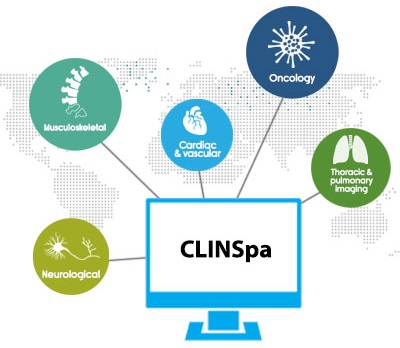
Our therapeutic expertise includes Oncology, musculoskeletal, central nervous system, neuro-oncology, neuro radiology, cardiac and vascular, thoracic and pulmonary imaging.
1. Oncology
Oncology trial is currently ICL’s fastest growing therapeutic area with over 50% of engagements in oncology trials. Experienced in large cancer trials with irRC and RECIST 1.1 protocols. Recently, RECIST and RECIST 1.1 were done together in the same trial.
2. Neurological
Strategic and practical support from experts that will boost your trial process and simplify your product development process. Our fellowship-trained neuroradiologists bring in a subspecialty level experience on neurodegenerative diseases such as Alzheimer’s Disease and Parkinson’s Disease, demyelinating disorders, stroke, epilepsy, neuro-oncology and post-traumatic disorders.
3. Cardiac and vascular
Our strength in Cardiac MR (CMR) image analysis, includes Anatomic Perfusion, Functional Imaging, Graft Patency and Stent Studies, as well as expertise in Nuclear Cardiology, further complements these services. A global team of in-house cardiovascular experts, who are highly-experienced in image analysis of clinical research and drug development
4. Thoracic and pulmonary imaging
We provide the highest level of expertise in the evaluation of chest radiographs and high-resolution CT of pulmonary disorders, including interstitial lung disease, chronic obstructive pulmonary disease, occupational lung diseases, pulmonary thromboembolism, bronchopulmonary infections and tumours.
CLINSpa- technology
A web-based platform that’s convenient and customizable designed tried and tested for an end to end management of imaging trial workflow. It allows you to customize workflows output templates thereby automating the process.
We aim to leverage our deep set expertise in technology and image interpretation service to accelerate drug development trials at the right speed.
To find out more about our Services, or how we can help you manage imaging requirements of your trial talk to us today..


