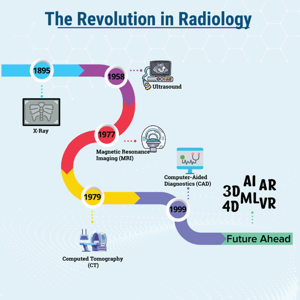

The past few decades have witnessed an amazing evolution in the arena of medical and clinical imaging. The field has come a long way enhancing precision and efficiency in diagnosis, triage, treatment, and research, revolutionizing the healthcare. In this blog, let us explore the captivating journey of medical and clinical imaging, following its development from infancy to the present day and moving ahead to what we can expect in the next decade.
The Beginning
The discovery of X-rays in 1895 by Wilhelm Conrad Roentgen marked a turning point in the development of medical imaging. This fundamental development enabled clinicians to investigate the internal organs of the human body without intrusive procedures. Both laymen and the medical community perceived this novel technology alike with amazement. Though primitive, the initial X-ray devices paved the way for further advancements in the discipline. The human curiosity and the insatiable appetite ushered advancements in medical imaging. Radiology made great strides in the 20th century.
Ultrasound (US) is a prominent imaging where high-frequency sound waves were used to create real-time images. In the second decade of the 20th century, US served as a beneficial therapeutic tool as a substitute for neurosurgery, rehabilitation, and the treatment of rheumatoid arthritis. Later, a doctor, Karl Theo Dussik, began researching US for medical imaging in the 1940s to identify the cerebral ventricles and locating brain tumors. In 1958, Gynecologist Ian Donald utilized US for the first time to examine the uterus, pelvis, and developing fetus. Later, Ultrasound became a crucial imaging in obstetrics, cardiology, and a number of other medical disciplines.
Computed Tomography (CT) scanning was invented by Sir Godfrey Hounsfield in 1972 and physicist Allan McLeod Cormack developed a similar system. Both jointly received a Nobel Prize in 1979. This technology marked a major breakthrough in medical imaging by delivering cross-sectional views of the body. Tumors, fractures, and other anomalies could be found with a level of accuracy never before possible. Later, CT perfusion and CT angiography revolutionized the field of stroke therapy.
Another frontier is magnetic resonance imaging (MRI) scanning which was developed by Dr Raymond Damadian in the 1977. It provided a non-invasive way of examining soft tissues, crucial for detecting diseases of the joints, spinal cord, and brain. It changed everything when detailed images were produced using strong magnets and radio waves.
In the 1990s, CAD (Computer-Aided Diagnostics) tools gained traction in mammography for breast cancer detection. In the 2000s, CAD expanded to lung radiology and other medical fields, improving diagnostic accuracy and efficiency.
Future Ahead
In the next decade, we can anticipate a cascade of advancements in the field of medical imaging. The integration of Computer-aided diagnosis (CAD) with Picture Archiving and Communication Systems (PACS) may improve lesion detection and differential diagnosis. Artificial intelligence (AI) and machine learning (ML) algorithms may accord with workflow platforms and help provide quicker and more precise visual interpretation.3D and 4D imaging and Functional imaging methods like positron emission tomography (PET) and functional MRI (fMRI) are the road to the future, which may offer a thorough understanding of intricate complex anatomical linkages and physiological processes. Compact and portable imaging devices, augmented reality (AR) and virtual reality (VR) are the waves of the future in technology-assisted medicine.
Adoption of novel medical imaging would accelerate the evolving field of precision medicine by delving deeper into the human body, providing explicit visual information on the anatomy and pathology of a patient. It would be possible to customize treatments based on each patient’s unique traits, improving therapy effectiveness and reducing side effects and enhancing patient care and management.


0 Comments